Saturday, September 30, 2006

Perfectionism and Anorexia

Beauty, fashion, and anorexia... when are they NOT in the news? But in the past few weeks, just like a model's hip bones, they've been even more prominent than usual.

Spain ban on skinny models shocks fashion world

By Andrew Hay Tue Sep 12, 11:30 AM ET

MADRID. (Reuters) - The world's first ban on overly thin models at a top-level fashion show in Madrid has caused outrage among modeling agencies and raised the prospect of restrictions at other catwalk pageants.

Madrid's fashion week has turned away underweight models after protests that young girls and women were trying to copy their rail-thin looks and developing eating disorders.


Firstview
Models Vlada Rosylakova (left)
and Natasha Poly (NYT).

Organizers say they want to project an image of beauty and health, rather than a waif-like, or heroin chic look.

But Cathy Gould, of New York's Elite modeling agency, said the fashion industry was being used as a scapegoat for illnesses like anorexia and bulimia.

Duh, Cathy, maybe that's because so many models are anorexic and bulimic! And just who, exactly, thinks this is attractive, anyway? You don't see boys obsessing over their I My Anorexic Girlfriend, Why Doesn't She Lose More Weight? MySpace pages! [all you pro-ana girls out there, please get some professional help! you have a serious psychiatric disorder.]

OK, what about all the girls with eating disorders who are not models or starlets? And what does this have to do with neuroscience, anyway??!

Just like nearly everything else these days, anorexia has been a target for fMRI research. Many of these studies have compared the neural responses to pictures of food in anorexic vs. control participants, and have found abnormally paltry responses in visual cortical areas and inferior parietal lobe (IPL) in the eating-disordered patients. At the same time, anorexic patients showed increased activity to pictures of food in the medial prefrontal cortex (PFC), relative to controls (Uher et al., 2004). The authors attributed the overactivation in medial PFC to a compulsiveness akin to OCD or drug addiction. A recent study manipulated whether the participants were hungry or satiated (Santel et al., 2006).
Santel S, Baving L, Krauel K, Munte TF, Rotte M. (2006). Hunger and satiety in anorexia nervosa: fMRI during cognitive processing of food pictures. Brain Res. Aug 15; [Epub ahead of print]

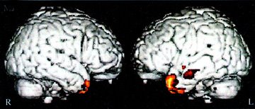
Neuroimaging studies of visually presented food stimuli in patients with anorexia nervosa have demonstrated decreased activations in inferior parietal and visual occipital areas, and increased frontal activations relative to healthy persons, but so far no inferences could be drawn with respect to the influence of hunger or satiety. Thirteen patients with AN and 10 healthy control subjects (aged 13-21) rated visual food and non-food stimuli for pleasantness during functional magnetic resonance imaging (fMRI) in a hungry and a satiated state. AN patients rated food as less pleasant than controls. When satiated, AN patients showed decreased activation in left inferior parietal cortex relative to controls. When hungry, AN patients displayed weaker activation of the right visual occipital cortex than healthy controls. Food stimuli during satiety compared with hunger were associated with stronger right occipital activation in patients and with stronger activation in left lateral orbitofrontal cortex, the middle portion of the right anterior cingulate, and left middle temporal gyrus in controls. The observed group differences in the fMRI activation to food pictures point to decreased food-related somatosensory processing in AN during satiety and to attentional mechanisms during hunger that might facilitate restricted eating in AN.
Most of the patients (n=9 out of 13) were inpatients, so
compliance with instructions was confirmed by clinical staff.
[What about the other four patients?? Oh, OK]
Of originally 14 patients, one patient was excluded because of non-compliance with instructions.
So when hungry, anorexic individuals may attempt to minimize the amount of attention granted to food stimuli, in order to bypass the intense signals telling them to EAT, EAT OR YOU'LL DIE! This isn't too surprising. The authors also hypothesize that in the rare instances when not hungry, decreased somatosensory processing (as indicated by decreased IPL activation) helps to limit the appeal of food, typically rated as disgusting by those with eating disorders.

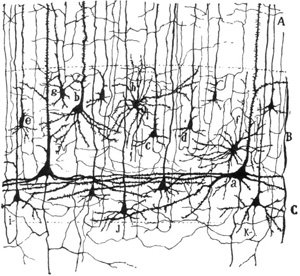
Finally, perfectionism has been strongly linked with anorexia. A recent EEG study reported that anorexics show an abnormal index of error processing. Specifically, the error-related negativity component (recorded when subjects make errors) was paradoxically reduced in the anorexia nervosa patients, who were much more focused on accurate performance than controls. Here, the authors suggested that medial PFC hypoactivity was responsible.
Pieters GL, de Bruijn ER, Maas Y, Hulstijn W, Vandereycken W, Peuskens J, Sabbe BG. (2006). Action monitoring and perfectionism in anorexia nervosa. Brain Cogn. Sep 6; [Epub ahead of print]

To study action monitoring in anorexia nervosa, behavioral and EEG measures were obtained in underweight anorexia nervosa patients (n=17) and matched healthy controls (n=19) while performing a speeded choice-reaction task. Our main measures of interest were questionnaire outcomes, reaction times, error rates, and the error-related negativity ERP component. Questionnaire and behavioral results indicated increased perfectionism in patients with anorexia nervosa. In line with their perfectionism and controlled response style patients made significantly less errors than controls. However, when controlling for this difference in error rates, the EEG results demonstrated a reduced error-related negativity in the patient group. These seemingly contradictory outcomes of improved performance and reduced error monitoring are discussed in relation with indications of anterior cingulate cortex hypoactivity in anorexia nervosa patients.
Fortunately, some in the fashion industry view anorexia as an illness, not as a beauty standard:
When Is Thin Too Thin?
By ERIC WILSON

. . .

"We are minutes away from a catastrophe," said David Bonnouvrier, the chief executive of DNA Models, which represents many of the top faces in the business. In an interview, Mr. Bonnouvrier said designers and model bookers were encouraging extreme thinness, so much so that several of the models he represents, when asked about their weight, have refused to seek medical attention for what are probable eating disorders.

"This goes against everything we stand for as an industry," Mr. Bonnouvrier said. "I am kicking and screaming about it now because this should be an industry of beauty and luxury, not famished-looking people that look pale and sick."
Reference
Uher R, Murphy T, Brammer MJ, Dalgleish T, Phillips ML, Ng VW, Andrew CM, Williams SC, Campbell IC, Treasure J. (2004). Medial prefrontal cortex activity associated with symptom provocation in eating disorders. Am J Psychiatry 161:1238-46.

OBJECTIVE: The authors sought to identify neural correlates of eating disorders in order to contribute to the debate on the genesis and classification of eating disorders and provide endophenotypes for genetic research. METHOD: Twenty-six female patients with eating disorders (10 with bulimia nervosa, 16 with anorexia nervosa) and 19 healthy female comparison subjects matched for age and education were presented with food and aversive emotional images while brain activity was recorded with functional magnetic resonance imaging. RESULTS: Women with eating disorders identified the food stimuli as threatening and disgusting. In response to these stimuli, the women with eating disorders had greater activation in the left medial orbitofrontal and anterior cingulate cortices and less activation in the lateral prefrontal cortex, inferior parietal lobule, and cerebellum, relative to the comparison group. In addition, women with bulimia nervosa had less activation in the lateral and apical prefrontal cortex, relative to the comparison group. Between-group differences in response to nonspecific emotional stimuli were found in the occipital cortex, parietal cortex, and cerebellum. CONCLUSIONS: A medial prefrontal response to symptom-provoking stimuli was identified as a common feature of anorexia and bulimia nervosa. This finding supports a conceptualization of eating disorders as being transdiagnostic at the neural level. The abnormal prefrontal reaction is associated with symptom-related material, whereas the occipital and cerebellar differences are nonspecific. An abnormal propensity to activate medial prefrontal circuits in response to inappropriate stimuli is common to eating, obsessive-compulsive, and addictive disorders and may account for the compulsive features of behavior in these conditions.

Subscribe to Post Comments [Atom]

Monday, September 25, 2006

Encephalon Seven

Encephalon 7th Edition now available at Omni Brain!

[or should I say Ensexalon?]
I figured we should start with the usual topics of the Omni Brain blog - sex, drugs, and rock and roll!


The Neurocritic's fictitious rendition of the stimuli from the study of Ponseti et al. (2006).

And still more sex on the brain! In The Frontal Cortex, Jonah Lehrer links to a Boston Globe article by Mark Liberman, who had previously written three excellent critiques of The Female Brain in Language Log.

Sex on the brain
By Mark Liberman

. . .

The Female Brain has made quite a splash since its publication last month, and this word-count claim is one of the most striking facts supporting her argument that the female brain is "a lean, mean communicating machine." The 20,000 vs. 7,000 numbers have been cited in reviews all over the world, from The New York Times to the Mumbai Mirror.

Since Brizendine is the director of a clinic at UCSF, one of the world's most important biomedical research institutions, and her book provides 90 pages of endnotes and references to back up 180 pages of text, I hoped it would finally give me a reliable source for this statistic.

The book's endnotes appear to attribute the numbers to a 1997 self-help book by Allan Pease and Allan Garner, Talk Language: How to Use Conversation for Profit and Pleasure. But Pease himself has presented several different word count numbers in other sources. In 2000, he published Why Men Don't Listen and Women Can't Read Maps (with Barbara Pease), which attributes to women "6,000-8,000 words," while men get "just 2,000-4,000 words." (They also offer daily counts for women's and men's "vocal sounds" and "facial expressions, head movements, and other body language signals"-but don't provide a source for any of the counts.) In a 2004 CNN interview, Allan Pease said that "women can speak 20,000 to 24,000 words a day versus a man's top end of 7,000 to 10,000."

And finally, Coturnix points to Sex, Science and Stereotypes, in which conservative political pundit David Brooks is taken to task, by Echidne of the Snakes, for his (mis)use of neuroscience research to "validate ancient stereotypes about the sexes" [don't worry if you don't get NYT Select, The Snake Goddess quotes the most relevant portions]. But of course, he's no neuroscientist; he relies on Louann Brizendine's book.

Subscribe to Post Comments [Atom]

Friday, September 22, 2006

Robot Skin

Continuing with the robot theme, the lowbrow science site LiveScience.com (infamous for sensationalistic headlines) carried a very funny article by Bill Christensen of Technovelgy.com,

Robots Get Soft, Human-Like Skin

If you hate being touched by your robot because of its cold metallic skin, take heart. Cosmetics manufacturer Kao Corporation and a Keio University research team led by robotics professor Takashi Maeno have developed artificial skin that feels just like yours. Or even softer.
Read the rest of the story.
According to the developers, ten out of twelve people agree that it feels like regular human skin. Machines also confirmed that the characteristics of the artificial skin werre similar to the skin of organic, natural humans.

Subscribe to Post Comments [Atom]

An "Endophenotype" For Sexual Orientation?

OR: The Neuroscience of Porn, according to The Frontal Cortex.

Wiktionary defines endophenotype as "any hereditary characteristic that is normally associated with some condition but is not a direct symptom of that condition."

Gottesman and Gould (2003) have this to say:
Endophenotypes, measurable components unseen by the unaided eye along the pathway between disease and distal genotype, have emerged as an important concept in the study of complex neuropsychiatric diseases. An endophenotype may be neurophysiological, biochemical, endocrinological, neuroanatomical, cognitive, or neuropsychological (including configured self-report data) in nature. Endophenotypes represent simpler clues to genetic underpinnings than the disease syndrome itself, promoting the view that psychiatric diagnoses can be decomposed or deconstructed, which can result in more straightforward—and successful—genetic analysis. However, to be most useful, endophenotypes for psychiatric disorders must meet certain criteria, including association with a candidate gene or gene region, heritability that is inferred from relative risk for the disorder in relatives, and disease association parameters. In addition to furthering genetic analysis, endophenotypes can clarify classification and diagnosis and foster the development of animal models. The authors discuss the etymology and strategy behind the use of endophenotypes in neuropsychiatric research and, more generally, in research on other diseases with complex genetics.

Gottesman II, Gould TD (2003). The endophenotype concept in psychiatry: Etymology and strategic intentions.
Am J Psychiatry 160:636-645.
However, for the study under discussion here, the "endophenotype" for sexual orientation is not diagnostic for a disorder of any sort, but merely for whether an individual (and his/her ventral premotor cortex) prefers to view pictures of the aroused genitals from one sex or the other. Do a Google seach for endophenotype, and the links are, in fact, mostly about disorders like schizophrenia, ADHD, autism, impulsivity, alcoholism, etc. Same for a PubMed search. Already we're dealing with a slightly loaded term, then, because the word endophenotype is associated with disorder.

But what about the pornography? Here it is,1 in NeuroImage:
Ponseti J, Bosinski HA, Wolff S, Peller M, Jansen O, Mehdorn HM, Buchel C, Siebner HR. A functional endophenotype for sexual orientation in humans. Neuroimage 2006 Sep 14; [Epub ahead of print]

Sexually arousing visual stimuli activate the human reward system and trigger sexual behavior. Here we performed event-related fMRI during visual processing of sexual core stimuli to pinpoint a neuronal correlate of sexual preference in humans. To dissociate gender of the stimulus from sexual preference, we studied male and female heterosexual and homosexual volunteers while they viewed sexual and nonsexual control stimuli. In contrast to previous work, we used core single-sex stimuli displaying male and female sexually aroused genitals. Since stimuli lacked any additional contextual information, they evoked no activity related to neuronal processing of faces, gestures or social interactions. Our prediction was that the sexual preference of the observer determines the neuronal responsiveness to pure male or female sexual stimuli in the human reward and motor system. Consistent with our prediction, the ventral striatum and the centromedian thalamus, showed a stronger neuronal response to preferred relative to non-preferred stimuli. Likewise, the ventral premotor cortex which is a key structure for imitative (mirror neurons) and tool-related (canonical neurons) actions showed a bilateral sexual preference-specific activation, suggesting that viewing sexually aroused genitals of the preferred sex triggers action representations of sexual behavior. The neuronal response of the ventral striatum, centromedian thalamus and ventral premotor cortex to preferred sexual stimuli was consistent across all groups. We propose that this invariant response pattern in core regions of the human reward and motor system represents a functional endophenotype for sexual orientation independent of the gender of the observer and gender of the stimulus.
OK, let's look at the study's German participants. There were 12 straight men, 12 straight women (0 or 1 on the Kinsey scale, meaning exclusively heterosexual or predominantly heterosexual) and 15 gay men, 14 gay women (Kinsey 6, meaning exclusively homosexual or 7... what's a Kinsey 7?? maybe they meant 5 and 6?). Anyway, those in the middle were excluded (sorry, all you bisexual Kinsey 2-4's out there, left out again). Below is the Kinsey Scale:

Rating

Description

0

Exclusively heterosexual

1

Predominantly heterosexual, only incidentally homosexual

2

Predominantly heterosexual, but more than incidentally homosexual

3

Equally heterosexual and homosexual

4

Predominantly homosexual, but more than incidentally heterosexual

5

Predominantly homosexual, only incidentally heterosexual

6

Exclusively homosexual

So in the scanner, the participants viewed, in random order, 30 pictures of female genitals, 30 pics of male genitals, and 30 neutral stimuli. The task was to press a button when an oddball target stimulus appeared (a green square). Immediately after the scan, the subjects rated the visual stimuli on arousal and valence.


Arousal ratings conformed more or less to the predicted pattern. In Fig. 1 above, the post-hoc comparisons between nonsexual stimuli (gray bars) and preferred sexual stimuli (red bars) are significant for all groups (albeit to a smaller degree for the straight women). In general, ratings for preferred vs. nonpreferred sexual stimuli were more discrepant in men than in women (regardless of orientation).

For the fMRI results, the authors defined 3 regions of interest, based on a priori hypotheses:

1. Centromedian Thalamus (and adjacent ventral striatum) - reward system [striatum, yes, but thalamus??]

2. Orbitofrontal Cortex - reward system

3. Ventral Premotor Cortex (PMv) - "motor representations of sexual behavior" [genital mirror neurons??]

Note that the amygdala was not among the ROIs, even though others have observed greater amygdala activity when viewing sexual stimuli compared to neutral stimuli (e.g., Hamann et al., 2004; see the Comments section of Sweat, Urine, and Sexual Orientation for a review of sex differences in brain activity when viewing porn clips). Come to think of it, the hypothalamus wasn't selected as an ROI, either. Hmmm, makes one wonder whether the ROIs were a posteriori...


Anyway, the red bars in the figure above (preferred sexual stimuli) depict greater activity than the yellow bars (nonpreferred sexual stimuli), which held for all four groups in thalamic regions, PMv, left superior parietal lobule [hey, what's that about??], but not orbitofrontal cortex.

Here's a thought. According to Wikipedia,
a mirror neuron is a neuron which fires both when an animal performs an action and when the animal observes the same action performed by another (especially conspecific) animal. Thus, the neuron "mirrors" the behavior of another animal, as though the observer were himself performing the action.
The Neurocritic is as skeptical as anyone about the mirror neuron craze, but if the PMv activity in this experiment is really imitative in nature, or even "empathetic" (instead of motor imagery or motor preparation), then wouldn't same-sex genitals elicit greater activity than opposite-sex genitals, regardless of sexual orientation?

Never mind.
The foci of premotor activation closely corresponded to the site that was found to be activated by observation of hand or mouth movements (Buccino et al., 2001) and by the observation of action-related objects (Grezes et al., 2003).

The observation of sexually aroused genitals of the preferred sex may trigger neuronal activity of mirror neurons or canonical neurons in the PMv. Sexually aroused genitals of the preferred sex are targets of manipulative actions during mating. Thus, in analogy to tools [NOTE: !!], these stimuli can be conceptualized as action-related objects which may activate canonical neurons in the PMv. Beyond its significance as a tool-like target, a sexually aroused genital belongs to an agent and indicates the motivational and bodily state of the agent (i.e., the readiness to mate).
[NOTE: So what would happen if you showed pictures of sex toys or sexbots or or... never mind.]

Ventral premotor mirror neurons may be involved in understanding the intention of the sexually preferred agent and activating a repertoire of volitional sexual actions.
[Hmm, seems like you could program a robot that way...]

But here's the bottom line:

We propose that this response reflect the activation of motor representations of manual or oral sexual behavior.
They really don't discuss the endophenotype idea much more, except to say,

We show a strong relation between the neuronal response and sexual orientation in distinct structures of the human reward and motor neuron system. ...the specific activity pattern induced by preferred sexual stimuli was remarkably invariant across heterosexual and homosexual males and females. In contrast to sexual preference, the sex of the stimulus and of the observer had no influence on the neuronal response to sexually aroused genitals. Therefore, we propose that the observed response pattern represents a functional endophenotype for sexual orientation in humans.

OK, then. Here's a teaser for a future post.
Moulier V, Mouras H, Pelegrini-Issac M, Glutron D, Rouxel R, Grandjean B, Bittoun J, Stoleru S. Neuroanatomical correlates of penile erection evoked by photographic stimuli in human males. Neuroimage 2006 Sep 6; [Epub ahead of print]

The objective of this study was to identify the cerebral correlates of the early phase, and of low to moderate levels, of penile tumescence using for the first time a volumetric measure of the penile response. We hypothesized that (i) regions whose response had been found correlated with circumferential penile responses in previous studies would be identified with volumetric plethysmography and (ii) that other brain regions, including the amygdalae, would be found using the more sensitive volumetric measurement. In ten healthy males, functional magnetic resonance imaging (fMRI) was used to study brain responses to sexually stimulating photographs and to various categories of control photographs. Both ratings of perceived erection and penile plethysmography demonstrated an erectile response to the presentation of sexually stimulating photographs. Regions where the BOLD signal was correlated with penile volumetric responses included the right medial prefrontal cortex, the right and left orbitofrontal cortices, the insulae, the paracentral lobules, the right ventral lateral thalamic nucleus, the right anterior cingulate cortex and regions involved in motor imagery and motor preparation (supplementary motor areas, left ventral premotor area). This study suggests that the development of low levels of penile tumescence in response to static sexual stimuli is controlled by a network of frontal, parietal, insular and cingulate cortical areas and that penile tumescence reciprocally induces activation in somatosensory regions of the brain.


1
The paper did not provide any examples of the stimuli used in the experiment. The reviewers apparently missed out on the opportunity to request that such images appear in an Appendix. But here's the verbal description (Ponseti et al., 2006).

First, sexual stimuli had to be specific to one or the other sex. Second, sexual stimuli should lack any further context information to avoid confounding brain activation related to neuronal processing of faces, gestures or social interactions. To meet these constraints we used photographs of naked male or female trunks displaying signs of genital arousal without showing the head or distal limbs. In addition we presented matched nonsexual photographs of the International Affective Picture System (IAPS) (Lang et al., 1997) as control stimuli. Male sexual stimuli showed an erected penis, female sexual stimuli showed details of the vulva (labia, clitoris, moist). The torso was sometimes visible together with the genitals, but the face or the limbs were never displayed on the photographs.

References

Buccino G, Binkofski F, Fink GR, Fadiga L, Fogassi L, Gallese V, Seitz RJ, Zilles K, Rizzolatti G, Freund HJ (2001). Action observation activates premotor and parietal areas in a somatotopic manner: an fMRI study. Eur. J. Neurosci. 13: 400-404.

Grezes J, Armony JL, Rowe J, Passingham RE (2003). Activations related to "mirror" and "canonical" neurones in the human brain: an fMRI study. NeuroImage 18: 928-937.

Hamann S, Herman RA, Nolan CL, Wallen K. (2004). Men and women differ in amygdala response to visual sexual stimuli. Nat Neurosci. 7: 411-6.

Subscribe to Post Comments [Atom]

Friday, September 15, 2006

Out And About

To avoid discovery I stay on the run. To discover things for myself I stay on the run.

--Jeanette Winterson, The PowerBook


The Neurocritic was very busy with one deadline after another, then went missing. Rumor has it that the semi-regular blogging schedule will resume shortly.

Subscribe to Post Comments [Atom]

Thursday, September 07, 2006

I Married A Robot

Metal Men No. 17
December 1965 / January 1966
Cover: Metal Men trapped in web as Doc Magnus gives wedding ring to Black Widow Spider robot
Story: "I Married a Robot" (25 pages)
Editor, writer: Robert Kanigher
Penciller: Ross Andru
Inker: Mike Esposito
Feature Characters: Metal Men (Gold, Lead, Iron, Mercury, Tin, Platinum (Tina), Nameless)
Supporting Character: Doc Magnus

Intro: Vilma (only appearance)
Villain: A Black Widow Spider robot, male spider robots (first and only appearance for all)
Synopsis: While Tina is trying valiantly to conquer Doc's heart again, a horde of robot spiders invades Earth, and their queen hypnotizes Doc into agreeing to be her mate.

Steve, you've done it again! Omni Brain has a link to an article on Marriage and Sex With Robots (An Ethical Perspective, at that), by David Levy. And no, I will not send you directly there, because the site is most definitely NOT WORK SAFE! So go to Omni Brain instead.

But if you manage to read the article (and believe me, I actually copied it into a Word document so I wouldn't have to view the ads), you'll find that there's actually nothing conceptually new there, just improvements in technology for more life-like sex toys... er uh, sexbots. Or, um, spousebots. And a discussion of the ethical implications for humans and robots alike. [John Searle! John Searle!] A prediction from David Levy:
Social change nowadays happens at a very much faster rate than it did in the nineteenth and most of the twentieth century, as a result of which I expect marriage with robots to be legalized in some countries by mid-century, with the state of Massachussets most likely being the first jurisdiction where a legally accepted human-robot marriage takes place. The prediction regarding Massachusetts is not only because of its comparatively liberal views to same-sex marriage but also because it is the home of countless high-tech companies and academic institutions working in the field of Artificial Intelligence.
And how do you Massachsetts liberals feel about human-robot marriage? [NOTE: the search term "robot marriage" did not appear on the aforementioned blog.]


Metal Men back story:

Showcase No. 37
March-April 1964
Cover: Metal Men vs. radioactive "manta-ray"
Story: "The Flaming Doom" (25 pages)
Editor, writer: Robert Kanigher
Penciller: Ross Andru
Inker: Mike Esposito
Feature Characters: Metal Men (Gold, Lead, Iron, Mercury, Tin, Platinum (Tina); a band of robots; first appearance for all; origin)
Supporting Characters: Dr. William (Will) Magnus (first appearance; last chronological appearance in flashback in BRAVE AND THE BOLD #55; full first name revealed in METAL MEN #47), Col. Henry Caspar (first appearance; last name spelled "Casper" in this story)
Intro: Amos J. Talbot, Al, Skipper (only appearance for all)
Villain: A manta ray-like radioactive creature (first appearance; dies in this story)
Synopsis: Dr. Will Magnus, a noted scientist, creates a platinum robot named Tina who displays human emotions and a body she can elongate and shape at will. Then, when a giant, flying, radioactive manta-ray creature endangers the Earth, he creates male robots from Iron, Tin, Gold, Mercury, and Lead, and sends them all against the monster as the Metal Men.


BREEDERS | Metal Man Lyrics
I don't know how old I was
But it was a '65 pickup
I was lying on the ground
With flat iron bars over my head
One silver drop is all you need
To put a hole in your head at 2000 degrees
That's right man, you be the Metal Man
At 2000 degrees
That's hot
that's hot

Etc.

Subscribe to Post Comments [Atom]

Wednesday, September 06, 2006

The EEG Mixer

Since Omni Brain has a post about the wonderfully enticing BrainBar, what's a bar and EEG without the requisite Electroencephalogram Cocktail Party??

The Science of Love

In Joyce Draganosky's
The Science of Love, the battle between reason and emotion takes center stage. A professor, who believes she has found a way of determining scientifically whether someone is in love, clashes with her department chair, a woman who thinks love and attraction are far too complex to be mapped according to the certainties of science.

Looks like a must-see short film if it ever comes to a city near you. But anyone can check out the clip on the web site above, which includes the hilarious "Electroencephalogram Cocktail Party" OR "The EEG Mixer"!! We can certainly forgive the confusion of fMRI and EEG results for artistic sake (and comic effect) here. The director seems to have modeled her lead character after Helen Fisher, even down to the combination of evolutionary anthropology with brain imaging. Joy Hirsh at Columbia was a scientific consultant.

But back to real life (Touching the Invisible is an art exhibition currently appearing in Stockholm, then in an unnamed locale in Canada).
Brainbar is a bar which serves you the drink you deserve. According to your state of mind at the time of ordering the drink, your brainwaves determine the metabolism between the machine and your own body.
The guest places the headband with biosensors on the forehead and then places the glass in the cylinder on the bar’s front side, to begin the reading. The biosensors then measure the state of the guest’s brain activity levels – shown with a flashing red light. The bar then administers the proper fluids for the guest’s psyche from the eight bottles and when the lamp flashes green the guest is allowed to remove the glass and enjoy the drink. The person can then join the party, confident that the fluids reacting with the internal metabolism are going to enhance the experience of that particular social event.

Although it's too late for this year's SFN meeting in Atlanta, perhaps the Behavioral and Cognitive Neuroscience Social should plan now for their 2007 event in San Diego (Nov. 3-7)!!

Subscribe to Post Comments [Atom]

They Found It!

Finally! Was it lost? What is it? Why, it's the brain's concept control core!

But I thought they found it in 1999 or 2000 (Mummery, Patterson, et al.).


Scientists identify brain's concept control core

By Jeremy Lovell

NORWICH (Reuters) - Scientists believe they may have finally identified the part of the brain that deals with the critical issue of matching words to everyday objects.

Using brain scans of people suffering from Semantic Dementia -- the second most common form of dementia after Alzheimer's disease in people under 65 -- they have found that the front end of the temporal lobe seems to be crucial to conceptual application.

"People have been talking about how the brain encodes concepts for 150 years. We believe we have found it," Matthew Lambon-Ralph of Manchester University told the annual meeting of the British Association for the Advancement of Science on Wednesday.
To be fair, Lambon-Ralph and his colleagues have done excellent work on semantic dementia for a long time (see refs). He's not responsible for the Reuters article. Here's a better quote:
"I have always been fascinated about how the brain gives meaning. We have made some strides toward that," he added.

Regions of significantly different grey matter density in a
semantic dementia patient (Mummery et al., 1999).


Then the science writer backtracks from the decisive headline (i.e., problem solved), which may not be his fault, and from the first sentence in the article:
However, much remains to be discovered about this particular brain function.

Not only do scientists not know which of the two temporal lobes is involved, they also do not know if other parts of the brain are also part of the conceptual process.

Only when they have discovered the answers to those crucial questions will they be able to work out how they can start to develop therapies to counteract the destructive effects of the progressive disease that eventually kills.
SUMMARY from The Neurocritic: Another case of "nice finding, but not a topic for the popular press to shout about."


References

Davies RR, Hodges JR, Kril JJ, Patterson K, Halliday GM, Xuereb JH. (2005). The pathological basis of semantic dementia. Brain 128:1984-95.

Lambon Ralph MA, Graham KS, Patterson K, Hodges JR. (1999). Is a picture worth a thousand words? Evidence from concept definitions by patients with semantic dementia. Brain Lang 70:309-35.

Jefferies E, Lambon Ralph MA. (2006). Semantic impairment in stroke aphasia versus semantic dementia: a case-series comparison. Brain 129:2132-47.

Mummery CJ, Patterson K, Price CJ, Ashburner J, Frackowiak RS, Hodges JR. (2000). A voxel-based morphometry study of semantic dementia: relationship between temporal lobe atrophy and semantic memory. Ann Neurol 47:36-45.

Mummery CJ, Patterson K, Wise RJ, Vandenbergh R, Price CJ, Hodges JR. (1999). Disrupted temporal lobe connections in semantic dementia. Brain 122:61-73.

Rogers TT, Lambon Ralph MA, Garrard P, Bozeat S, McClelland JL, Hodges JR, Patterson K. (2004). Structure and deterioration of semantic memory: a neuropsychological and computational investigation. Psychol Rev 111:205-35.

Williams GB, Nestor PJ, Hodges JR. (2005). Neural correlates of semantic and behavioural deficits in frontotemporal dementia. Neuroimage 24:1042-51.

Subscribe to Post Comments [Atom]

Monday, September 04, 2006

The Synapse, Vol. 1, issue 6


Représentation des neurones du cortex cérébral par Cajal


An exciting new issue of The Synapse is available now at The Mouse Trap!

Subscribe to Post Comments [Atom]

eXTReMe Tracker